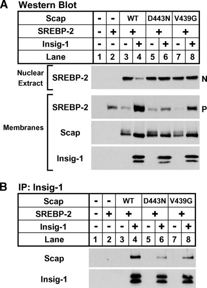
Fig. 5.
Hamster Scap mutant V439G mediates insulin-induced gene (Insig)-resistant SREBP activation and is defective in Insig binding. On day 0, SRD-13A cells were set up in medium A at 9 × 10⁵ cells per 10 cm dish. On day 2, cells were transfected in medium B with 4 μg of pTK-HSV-SREBP-2, 0 or 300 ng of pCMV-Insig-1-Myc, and 1 μg of the indicated pCMV-Scap plasmid. Three hours after transfection, cells were refed with medium B containing 50 μM compactin and 50 μM mevalonate and incubated for 18 hours. N-acetyl-leucyl-leucyl-norleucinal (ALLN) (25 μg/ml) was added to each dish 2 hours prior to harvest. Cells were fractionated as described in Experimental Procedures, and membranes were divided into two aliquots and used for both Western blot analysis (A) and immunoprecipitation (B). A: Nuclear extracts (0.3 dish of cells) and membranes (0.25 dish of cells) were subjected to Western blot analysis with anti-herpes simplex virus (HSV) IgG (SREBP-2), anti-Scap IgG 9D5, or anti-Myc IgG 9E10 (Insig-1). N and P denote the nuclear and precursor forms of SREBP-2, respectively. B: Digitonin-solubilized membranes (0.5 dish of cells) were subjected to immunoprecipitation (IP) with anti-Myc IgG 9E10 and protein A-agarose. Bound fractions were subjected to Western blot analysis with polyclonal anti-Myc IgG (Insig-1) or anti-Scap IgG R139.

