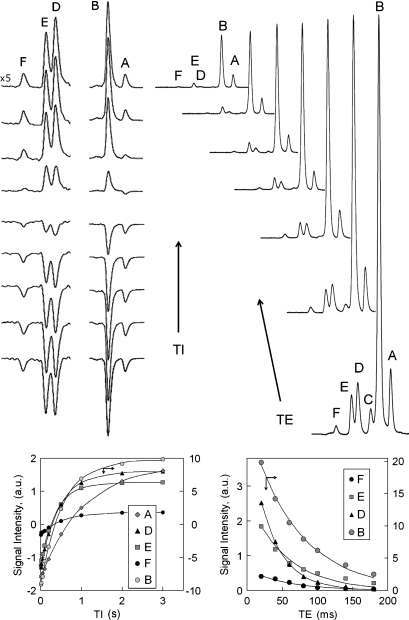
Fig. 3.
Inversion-recovery measurement of T1 (left panel) and echo time (TE)-dependence measurement of T2 (right panel) from subcutaneous fat tissues of a 34 year-old healthy male at 7T. The inversion bandwidth (BW) was set to span resonances A and B (middle stack), or resonances C, D, E, and F (left-most stack). The inversion delay times for the given spectra are 10, 20, 50, 100, 200, 500, 1,000, 2,000, and 3,000 ms with a constant repetition time (TR) of 7 s and TE of 40 ms (left panel). The echo times for the T2 measurements are 20, 40, 60, 80, 100, 140, and 180 ms (right panel).

