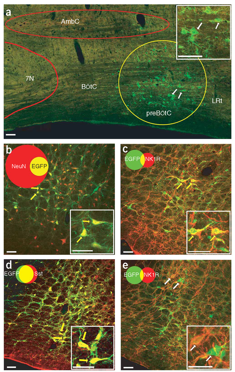
Figure 1.
preBötC neurons infected by AAV2 containing a Syn or Sst promoter. (a) EGFP expression (green) in adult rat preBötC (yellow circle) 3 weeks after microinjection of Syn-EGFP AAV2. 7N, facial nucleus; AmbC, ambiguous nucleus compact; BötC, Bötzinger Complex; LRt, Lateral reticular nucleus. (b) Syn-AlstR-EGFP AAV2–infected preBötC cells (green) were NeuN immunoreactive (red); that is, they were neurons. (c) Some Syn-AlstR-EGFP AAV2–infected preBötC neurons (green) were NK1R immunoreactive (red). (d) Sst-EGFP AAV2–infected preBötC neurons (green) were Sst immunoreactive (red). (e) Small portion of Sst-AlstR-EGFP AAV2–infected preBötC neurons (green) were NK1R immunoreactive (red). Images were obtained using a confocal microscope. Insets are high magnification of indicated neurons. Venn diagrams represent the relative number of neurons and the overlap of the relevant markers (b–e) normalized to NeuN immunoreactivity (n ≈ 3,000 neurons on each side of the preBötC). Yellow arrows represent neurons coexpressing and white arrows represent neurons not coexpressing the relevant markers. Scale bars represent 50 µm.

