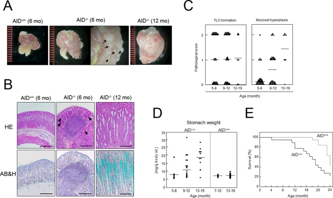
Figure 1. AID−/− mice spontaneously developed gastritis.
(A) Stomachs of AID+/+ and AID−/− mice were cut longitudinally to expose the gastric mucosae. Inside-out tissues were examined by stereomicroscopy. A high magnification view of the mucosal surfaces revealed the presence of a number of nodule-like structures in the AID−/− mouse stomach (arrows). A representative sample from each group is shown. (B) Gastric tissue sections were stained with H&E for histological examination, or with Alcian blue-hematoxylin (AB&H) for detection of mucin-producing cells (shown as turquoise). Scale bars: 100 µm. (C) Gastric tissues of AID−/− mice at different ages were analyzed for development of ectopic lymphoid follicles and mucosal hyperplasia. The diagnostic criteria are described in Supplemental Table S1 online. (D) Stomach weights of AID+/+ and AID−/− mice were measured and the values were normalized to the body weight of each mouse. (E) Kaplan-Meier survival curves of AID−/− (solid line, n = 18) and AID+/+ (dotted line, n = 19) mice revealed significant differences in survival (P<0.05).

