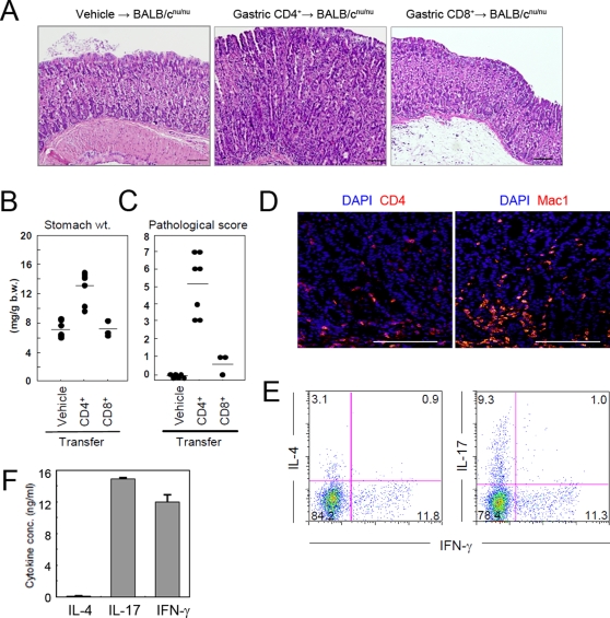
Figure 8. CD4+ T cells contribute to the development of gastritis in AID−/− mice.
Gastric mucosa-infiltrating CD4+ T cells or CD8+ T cells were isolated from 12 month old AID−/− mice and were adoptively transferred into BALB/cnu/nu mice. After 3 months, the stomachs of recipient mice were removed and subjected to the following experiments. (A) Gastric tissue sections were stained with H&E for histological examination. (B) Stomach weights were normalized to body weight of each mouse. (C) Gastric tissue sections were examined by histology and assigned a pathological score. (D) Immunofluorescence staining revealed a massive infiltration of CD4+ and Mac1+ cells into the stomachs of BALB/cnu/nu mice that received CD4+ T cells by adoptive transfer. (E) Gastric CD3+CD4+ cells enriched with a MACS column were treated with PMA and Golgi-stop for 6 hr and analyzed for cytokine production by intracellular staining. (F) CD4+ T cells recovered from gastric tissues of CD4+ T-cell-transferred BALB/cnu/nu mice were stimulated with anti-CD3 and anti-CD28 mAbs for 48 h. Cytokine concentrations in the culture supernatants were measured by CBA and ELISA. Scale bars: 100 µm.

