
Figure 4. In vitro suppression assays.
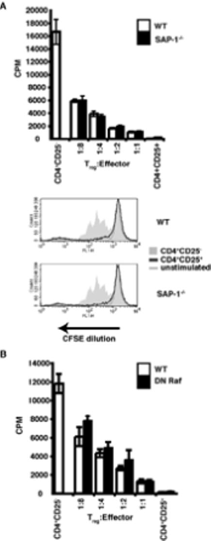
(A) Thymidine incorporation assays were used to assess in vitro suppressive function at a range of effector:Treg ratios. Thymic CD4+CD25- effectors and Tregs were used. +/+, wild-type; -/-, SAP-1 deficient, white bars, wild-type; black bars, SAP-1-/-. CFSE suppression assays performed at a 1:1 ratio of Teffector:Treg. Grey shaded area, activated effectors at 72hrs; dark grey line, 1:1 ratio of Teffectors:Tregs; light grey line, unstimulated effectors. Top panel wild-type thymocytes, Bottom panel SAP-1-/- thymocytes (B) Suppression assay, assessed by thymidine incoportaion after 72hrs. White bars, wild-type; black bars, DN Raf T cells. CD4+CD25- effectors and Tregs were taken from lymph nodes.