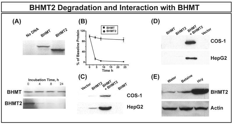
Figure 5.
BHMT2 translation, degradation and interaction with BHMT. (A) Human BHMT and BHMT2 in vitro translation in a rabbit reticulocyte lysate (RRL) is shown in the top panel. Representative autoradiographs for 35S-methionine radioactively-labeled BHMT and BHMT2 at different time points in degradation experiments are shown in the bottom panel. The data shown are representative of three independent experiments. (B) BHMT and BHMT2 protein remaining at each time point in the RRL degradation study, expressed as percentages of the basal value. Each point is the mean ± SEM for 3 independent experiments. BHMT2 differed significantly from BHMT at each point (p < 0.0001). (C) Immunoblot analysis of HA-tagged BHMT2 expressed in COS-1 and HepG2 cells either together with BHMT or alone. Western blotting was performed with anti-HA antibody. Samples were loaded on the SDS-PAGE gels based on cotransfected β-galactosidase activity to correct for possible variation in transfection efficiency. (D) BHMT2 and BHMT interaction. HA-tagged BHMT2 was transfected or cotransfected with BHMT into COS-1 and HepG2 cells. IP was performed with equal quantities of total protein and anti-HA antibody, followed by immunoblot analysis with anti-BHMT antibody. Results shown are representative of 3 independent experiments. (E) HA-tagged BHMT2 expressed in HEK293T cells was stabilized in the presence of 1 mM homocysteine. Cells expressing HA-tagged BHMT2 were cultured with 1 mM homocysteine (Hcy), 1 mM betaine or water for 24 h. Western blot analysis was performed with anti-HA antibody, and actin was used as a control for loading.

