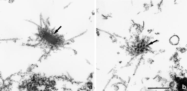
Figure 4.
Interphase centrosome. (a) Microtubules are originating from the corona (arrow) surrounding the rectangular core of the centrosome. Note the layered composition of the core. (b) Grazing section of the corona, showing the more or less regular arrangement of electron-dense nodules (arrow). Scale bar, 0.5 μm. The electron micrographs of this figure as well as Figure 5 are from cells lysed during fixation according to the protocol described in MATERIALS AND METHODS.

