Figure 3.
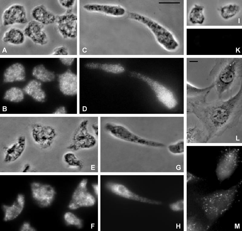
(A–K) GRP125 and its homologue localize to vesicular compartments in Dictyostelium cells. Corresponding phase-contrast micrographs (A, C, E, G, and I) and fluorescence images (B, D, F, H, and K) at different developmental stages are shown. Bar, 10 μm. (A–D) Localization of GRP125 in growth phase (A and B) and aggregation-competent (C and D) D. discoideum AX2 cells. Fixation was achieved using cold methanol; specimens were processed for indirect immunofluorescence labeling of GRP125 using the polyclonal anti-GRP125/S2S3 antibody followed by Cy3-labeled goat anti-rabbit IgG. (E–H) Immunofluorescence localization of both GRP125 and its homologue in growth phase (E and F) and aggregation-competent (G and H) Dictyostelium cells. Cells were fixed with picric acid/formaldehyde/70% ethanol. Labeling of both GRP125 and its homologue was performed using the polyclonal anti-GRP125/S3S4 antibody followed by the Cy3-labeled goat-anti rabbit IgG. (I and K) Background fluorescence using only the secondary Cy3 labeled goat-anti rabbit IgG antibody. (L and M) Incorporation of the recombinant rhodamine-labeled GRP125/S3S4 polypeptide to vesicle-like subcellular compartments after microinjection into living NIH/3T3 fibroblasts. (M) Fluorescence; (L) corresponding phase-contrast image. Bar, 10 μm.

