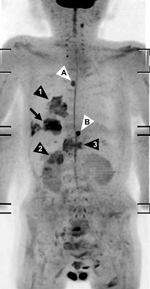
Fig. 11.
A 78-year-old man with metastatic lung cancer and two benign lesions visualized with DWIBS. Coronal maximum intensity projection DWIBS image shows an area with restricted diffusion in the right lower lobe, consistent with lung cancer (arrow). In addition, DWIBS image shows ipsilateral hilar lymph node metastasis (arrowhead 1), retroperitoneal/adrenal metastasis (arrowhead 2), and vertebral metastasis (arrowhead 3). DWIBS image also shows restricted diffusion in a thoracic vertebra (arrowhead A). T1- and T2-weighted images (not shown), however, indicate this “lesion” to be a (benign) cavernous hemangioma, instead of a vertebral metastasis. Furthermore, DWIBS image shows a well-circumscribed area of restricted diffusion in the epigastric region (arrowhead B); physical examination reveals a (benign) sebaceous cyst to be responsible for this area of restricted diffusion

