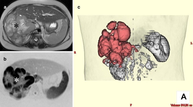
Fig. 9.
Volume measurement of a primary hepatic diffuse large B-cell lymphoma in a 10-year-old girl using DWIBS. a Axial respiratory-gated T2-weighted image shows a bulky mass occupying most of the right lobe of the liver (asterisk). b Axial DWIBS image (inverted black-and-white gray scale) at the same level clearly visualizes the tumor with excellent contrast between the tumor and surrounding tissue (asterisk). c Volume-rendered image using thin slice (4 mm, no gap) DWIBS dataset shows the three-dimensional shape of the tumor (colored red) and allows for tumor volume measurement

