Figure 1.
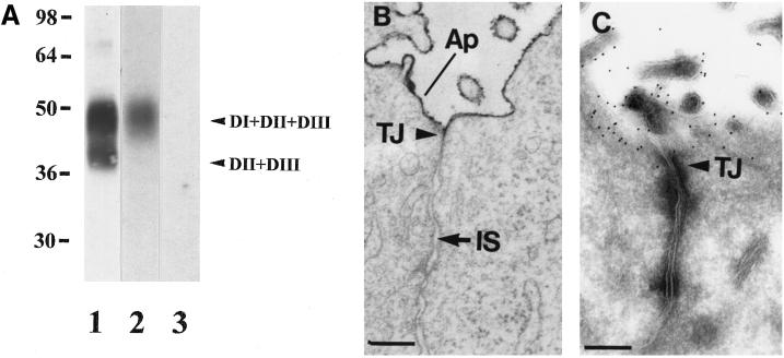
Transfected uPAR is expressed correctly as an apically sorted, GPI-anchored protein and specifically binds DFP-uPA. (A) TX-100 (1%) extracts from transfected MDCK cells were resolved by 10% SDS-PAGE and transferred to nitrocellulose membranes for Western blotting with polyclonal rabbit antibodies that recognize full-length (DI+DII+DIII) and cleaved (DII+DIII) uPAR (lane 1) or ligand blotting with 10 pM [125I]-DFP-uPA alone (lane 2) or 100 nM unlabeled ligand (lane 3). (B) MDCK cells grown to confluency for 4 d in plastic dishes were fixed in the presence of ruthenium red before embedding. Ruthenium red is restricted to the apical (Ap) cell surface and microvilli and excluded from the intercellular space (IS) by tight junctions (TJ). (C) Polarized MDCK cells cultured as in B were fixed and processed for cryoimmunogold labeling with uPAR antibodies followed by 10 nm colloidal gold–conjugated protein A. Labeling is confined to the apical domain by tight junctions (TJ). Bars, 100 nm.

