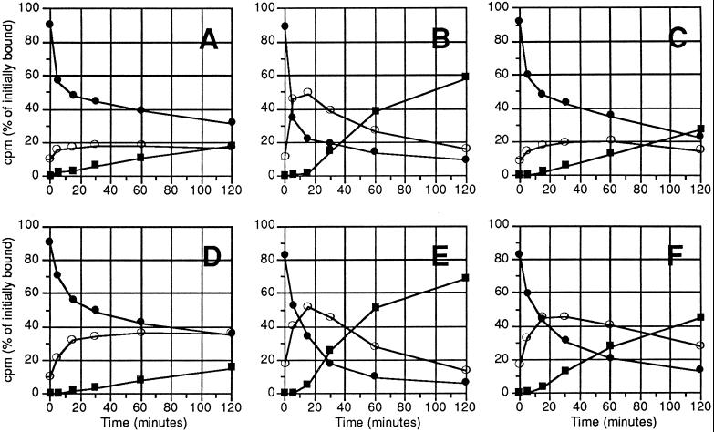
Figure 2.
Time course of internalization and degradation of DFP-uPA and uPA:PAI-1 in polarized and nonpolarized MDCK cells. Polarized monolayers cultured for 4 d (A–C) or nonpolarized MDCK cells cultured for 14–16 h (D–F) were incubated with 500 pM [125I]DFP-uPA (A and D) or [125I]uPA:PAI-1 without (B and E) or with (C and F) 100 nM RAP for 90 min at 4°C, washed, and then shifted to 37°C in the absence (B and E) or presence (C and F) of 100 nM RAP. At the times indicated the fraction of ligand present on the cell surface (closed circles) and in the cell lysate (open circles) or as degraded ligand in the medium (squares) was determined as described in MATERIALS AND METHODS and expressed as percentage of total cell-associated counts before shift to 37°C. A representative experiment of three to six independent ones is shown.

