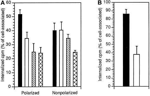
Figure 3.
Polarized and nonpolarized MDCK cells show differential sensitivity to inhibition of [125I]uPA:PAI-1 internalization by RAP and excess unlabeled uPA:PAI-1. (A) Polarized or nonpolarized MDCK cells were incubated with 500 pM [125I]uPA:PAI-1 for 90 min at 4°C. After washing cells were incubated for 10 min at 37°C in the absence (black bars) or presence of either a 200-fold molar excess of unlabeled ligand (white bars) or 100 nM RAP (hatched bars). For comparison, endocytosis of prebound [125I]ricin (200 ng/ml) was also determined (cross-hatched bars). Surface-associated and internalized counts were then determined, and results are expressed as internalization index (internalized counts per minute/total cell-associated counts per minute). (B) Polarized (black columns) or nonpolarized (white columns) MDCK cells were incubated with 1 nM [125I]RAP at 4°C and then chased for 7 min at 37°C before measurement of surface-associated and internalized ligand. Results are expressed as internalization index. In both A and B, mean and SD of three independent experiments in duplicate are shown.

