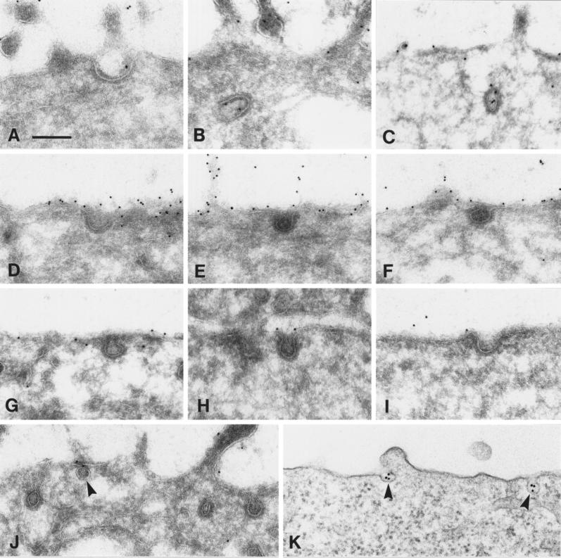
Figure 4.
In nonpolarized cells uPAR is excluded from clathrin-coated pits and localizes in caveolae only in the presence of cross-linking antibodies. (A–C) Polarized MDCK cells were incubated with 10 nM uPA:PAI-1 at 4°C and then chased for 10 min at 37°C. Subsequently, cryosections were labeled with anti-uPAR rabbit antibodies followed by protein A-gold (10 nm). Representative images of coated pits containing uPAR are shown. (D–F) Cryosections derived from an MDCK clone (clone 8.1) expressing uPAR at very high levels. Cells were fixed at subconfluency (in the absence of ligand) and labeled for uPAR. (G–J) Nonpolarized MDCK cells (clone 3.2) were challenged with uPA:PAI-1, and cryosections were labeled for uPAR. Note that in both types of experiment (D–J), uPAR was largely excluded from coated pits. Moreover, uPAR in general was also excluded from caveolar profiles (J, arrowhead). (K) Epon section of nonpolarized MDCK cells, which were incubated with anti-uPAR rabbit antibodies (1.5 μg/ml) followed by 10 nm of gold-conjugated goat anti-rabbit antibodies before fixation and processing for electron microscopy. Note clustering of uPAR in caveolae (arrowheads). Bar, 200 nm.

