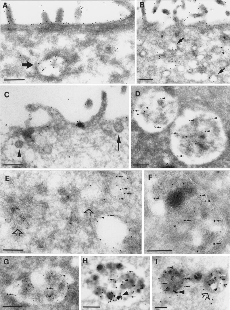
Figure 9.
uPA:PAI-1 causes partial redistribution of uPAR to late endosomes and lysosomes in nonpolarized MDCK cells. (A and B) Polarized MDCK cells; (C–I) nonpolarized MDCK cells. Cells internalized prebound uPA:PAI-1 for 150 min at 37°C (A–G) or, alternatively, were loaded with cationized 20-nm gold for 5 h, chased in DMEM-BSA alone for 3 h, and then incubated with 50 nM uPA:PAI-1 for 3 h in DMEM-BSA containing 10 μg/ml cycloheximide (G and H). Ultrathin cryosections were labeled with anti-uPAR antibodies followed by 10 nm gold-conjugated protein A (A–C, H, and I) or double labeled with secondary anti-MPR (D and E) or anti-LAMP (F and G) antibodies followed by 15 nm gold-conjugated protein A. (A and B) In polarized MDCK cells uPAR was mainly expressed on the cell surface but was also present in multivesicular bodies (A, large arrow) or in small apical endosomes (B, arrows). (C) Cell surface expression of uPAR in nonpolarized MDCK cells. The arrow points to a clathrin-coated pit; the arrowhead points to an apparent clathrin-coated vesicle. (D) Colocalization of uPAR (arrowheads) and MPR (arrows) in large endosomes with internal membrane structures. (E) uPAR (arrowheads) and MPR (arrows) colocalize in endosomes, but tubulovesicular structures (open arrows) show exclusive labeling for uPAR. (F and G) Colocalization of uPAR (arrowheads) and LAMP-1 (arrows) in dense lysosomal compartments. (H and I) uPAR (arrows) and cationized 20-nm gold particles (arrowheads) colocalize in compact lysosomes. Note the presence of aggregated, cationized gold particles (large arrowheads). (I) The open arrow points to a lysosome with many cationized-gold clusters but no uPAR. Bars, 250 nm.

