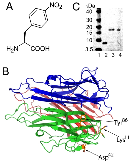
Fig. 1.
Incorporation of pNO2Phe into mTNF-α. (A) Structure of pNO2Phe. (B) X-ray crystal structure of mTNF-α trimer with Tyr-86, Asp-42, and Lys-11 indicated (PDB ID code 2TNF). (C) Expression of the Tyr86 amber mutant of mTNF-α in the absence (lane 2) and presence (lane 3) of 1 mM pNO2Phe with the pNO2Phe-specific mutRNACUA/aminoacyl-tRNA synthetase pair. Protein samples were purified by Ni-NTA affinity column under denaturing conditions and analyzed by SDS/PAGE with SimplyBlue staining. Lane 4 contains WT mTNF-α, and lane 1 is a molecular mass standard.

