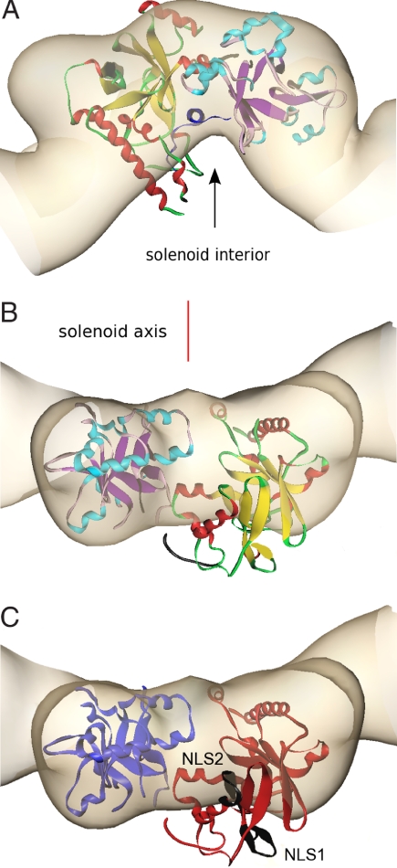
Fig. 3.
Alignment of the crystal structure of VirE1–VirE2 complex with the VirE2 envelope obtained by EM in the presence of ssDNA, represented at the same length scales. The crystal structure of VirE2 (colors as in Fig. 2A), treated as a single rigid body, was manually aligned into the envelope of one repeat of VirE2 in the solenoid as determined by EM (beige) and subject to constraints as described in the text. [Figures created with Amira (Mercury Computer Systems)]. (A) The view down the solenoid axis. For alignment purposes, VirE1 (dark blue) was introduced as shown facing the solenoid interior (black arrow), although it is in fact absent from the VirE2 complex with ssDNA. (B) Rotation of view A by 90° perpendicular to the solenoid axis (red line), showing one VirE2 repeat viewed from the exterior of the solenoid. It is clear that, although one domain of VirE2 neatly fits the envelope, the other protrudes from it (the interdomain flexible linker is shown in black). (C) Indication of the two NLS sequences (black) facing the exterior of the solenoid. The orientation is as in B, and the color scheme is simplified for clarity (N-terminal domain, red; C-terminal domain, blue).

