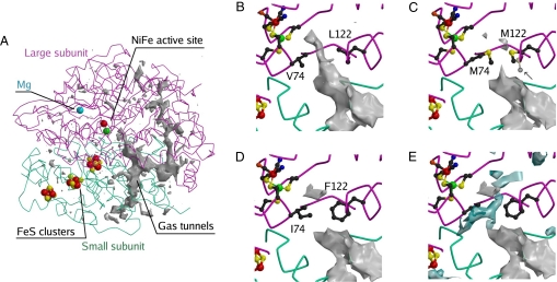
Fig. 1.
Structural models of the three enzymes. A gives an overview of the tunnel network; B is a closeup of the tunnel near the active site in the WT. C, D, and E are closeups of the MM and FI mutants, as indicated. In C, an arrow points to the second conformation of M122. A conserved hydrophilic cavity is shown in blue in E.

