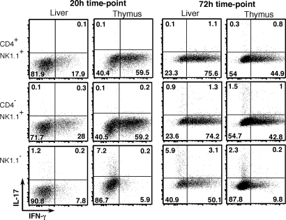
Fig. 2.
Costaining of NKT cell subsets for IFN-γ and IL-17. NKT cell subsets were isolated and stimulated on anti-CD3– and anti-CD28–coated plates as described in Fig. 1. GolgiStopTM was added to cultures 4 h before the indicated time point, and cells were stained for intracellular IFN-γ-APC and IL-17-PE. Numbers in each quadrant represent the percentage of cells in that quadrant, and results are representative of two independent experiments.

