Fig. 1.
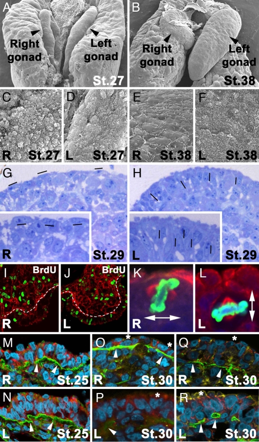
Asymmetrical differences in female chick gonad embryogenesis. (A and B) Scanning electron micrographs at stage 27 (A) and stage 38 (B) HH. (C–F) Right (C and E) and left (D and F) surface of symmetrical stage 27 HH (C and D) and asymmetrical stage 38 HH (E and F) gonads. Note the loss of a globular shape and pilli distribution on the right gonad (C and E). (G and H) Histological sections of right (G) and left (H) gonads at stage 29 HH stained with Toluidine blue illustrating condense packaging of nuclei in the left gonad cortex (H). Magnifications of right and left cortex are evident in the Insets of G and H. Hyphens show the orientation of oval nuclei. (I and J) BrdU incorporation (green) in right (I) and left (J) gonad nuclei after a 1-h pulse at stage 28 HH. A white line marks the borders of the gonad cortex to compare differential proliferation. Basal lamina is evident by laminin immunostaining in red. (K and L) Representative mitotic cells for right (K) and left (L) gonads with perpendicular and parallel metaphase planes, respectively, at stage 27 HH (nuclei shown in blue, Phospho Histone H3 in green, and α-tubulin in red). (M–P) Immunohistochemistry detection for laminin (green) and β-catenin (red) at stage 25 HH (M and N) and 30 HH (O and P) in the right (M and O) and left (N and P) gonads. (Q and R) Immunostaining for laminin (green) and N-cadherin (red) at stage 30 HH in the right (Q) and left (R) gonads. In M–R, DNA is shown in cyan and actin is shown in yellow. At stage 30 HH, basal lamina continuity is maintained in the right gonad (O and Q) but not in the left gonad (P and R), compared with the symmetry detected in both gonads at stage 25 (M and N; white arrowheads in M–R point to basal lamina). Note a loss of the intense detection of β-catenin and N-cadherin in the apical cytocortex at stage 30 HH (asterisks) when morphological asymmetry between left and right gonads is evident (O–R).

