Fig. 3.
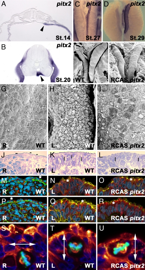
Pitx2c regulates gonad morphogenesis. (A and B) Histological sections showing pitx2c expression in left gonad primordia (black arrowhead) at stage 14 HH (A) and 20 HH (B). Left-sided pitx2c expression is detected before any overt difference is observed between the left and right gonads. Subsequently, expression is maintained through cortex asymmetric differentiation stages (stage 27 in C and stage 29 in D). Overgrowth of the right gonad 48 h after pitx2c overexpression (F) can be compared with a control stage 31 HH right gonad (E). (G–U) Phenotype of right gonads after pitx2c overexpression. (G, J, M, P, and S) Right gonads. (H, K, N, Q, and T) Left gonads. (I, L, O, R, and U) Right gonads after pitx2c overexpression. Scanning electron microscopy micrographs show patches with high pilli density in right gonads after pitx2c overexpression as observed for left wild-type gonads (I, compare with G and H). Histological sections show compactness of nuclei density and a shift to a perpendicular orientation on the right gonad after pitx2c overexpression (L, compared with J and K). Hyphens indicate nuclei orientation. Immunohistological sections show transformation of the right gonad to left phenotype after pitx2c overexpression with laminin and β-catenin (O, compare with M and N) and laminin and N-cadherin (R, compare with P and Q). Note the loss of basal lamina continuity, increase of nuclei packaging, and detection of N-cadherin, β-catenin, and actin in the external section of the cytoplasm after pitx2c overexpression (asterisks). In M–R, DNA is shown in cyan and actin is shown in yellow. (S–U) Cell division plane in pitx2c transformed (U) and in control (A/P-infected) right (S) and left (T) gonads (nuclei shown in blue, Phospho Histone H3 in green, and α-tubulin in red). pitx2c overexpression increases the percentage of parallel mitotic divisions in the right gonad. Double arrows point to the orientation of the mitotic spindle.

