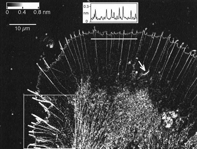
Figure 1.
Birefringent fine structure in the living growth cone of an Aplysia bag cell neuron. This retardance magnitude image, recorded with the Pol-Scope, shows the peripheral lamellar domain containing radially aligned birefringent fibers that end in filopodia at the leading edge of the growth cone. The graph in the top center shows the retardance magnitude, in nanometers, measured along a three-pixel-wide strip that is indicated by a horizontal white bar below the graph. The measured peak retardance in radial fibers can be interpreted in terms of the number of filaments in the fibers. Near the bottom, the image shows the central domain, which is filled with vesicles and highly birefringent fibers. On the bottom left, a region is outlined, and its image contrast is enhanced to show clearly the diffuse birefringent patches located mainly in the transition region between the thin lamellar and thicker central domain. The arrow near the center of the image points to a spontaneously formed intrapodium with its strongly birefringent tail. The image is one frame of a time-lapse record that reveals the architectural dynamics of the cytoskeleton including the formation of new filopodia and radial fibers and the continuous retrograde flow of birefringent elements in the peripheral domain. The top left corner of the image contains a gray horizontal bar with the retardance scale and a 10 μm scale bar.

