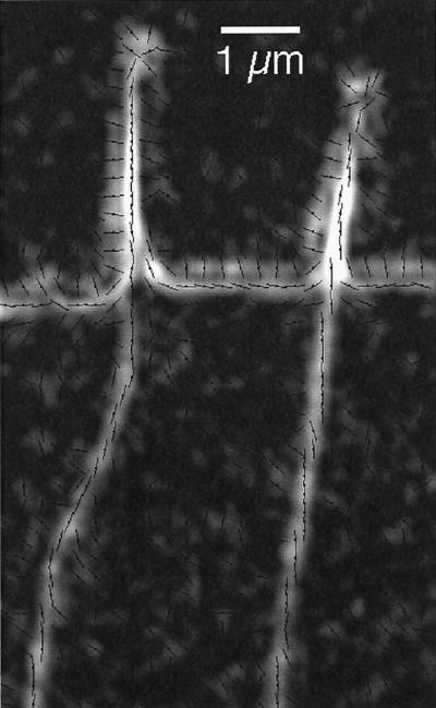
Figure 2.
Radial fibers and filopodia near the leading edge recorded by the Pol-Scope (highly enlarged with digital smoothing). The retardance magnitude image is overlaid by short lines indicating the orientations of the slow birefringence axis measured in each location. The birefringence of radial fibers and the cores of filopodia have slow axis orientations that are parallel to the fiber axis. The cell membrane at the leading edge is imaged as a birefringent double layer with mutually orthogonal slow axis orientations. The double layer does not represent the lipid bilayer but is mainly caused by the refractive index difference between the optically denser cytoplasm and the surrounding medium. The slow axis of the double layer is parallel to the interface at the high-refractive index side (cytoplasm) and perpendicular to the interface at the low-refractive index side (medium) (Oldenbourg, 1991). White corresponds to 0.34-nm retardance. Slow axis orientations are typically shown for every second pixel in the horizontal and vertical direction of the original video frame. Bar, 1 μm.

