
Figure 3. Aberrant hair follicle development and global expression of placode and hair shaft markers in activated β-catenin mutant embryos.
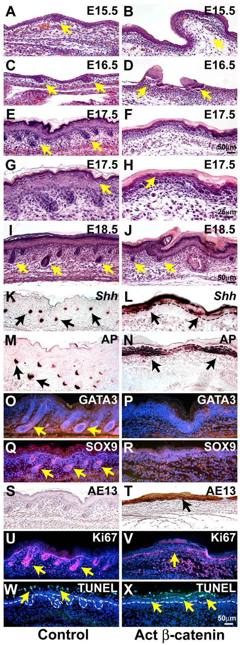
(A – J) Histological analysis of dorsal skin from control littermate and KRT14-Cre Ctnnb1(Ex3)fl/+ mutant embryos reveals failure of hair follicle downgrowth in mutants. Mutant skin displays evaginations (B, D, J) and fails to stratify normally (H). (G, H) are higher magnification views of (E, F) respectively. The placode marker Shh (in situ hybridization, brown) (K,L) and dermal condensate marker alkaline phosphatase (AP) (enzymatic assay, purple brown) (M,N) are expressed broadly in the mutant. Expression of the inner root sheath marker GATA3 (O,P) and the outer root sheath marker SOX9 (Q,R) (immunofluorescence, red) is reduced or absent, but hair shaft keratins (AE13 antibody, immunohistochemistry, brown) are ectopically expressed in mutant ectoderm (S, T). Proliferation is decreased (U,V) (Ki67 immunofluorescence, red) and the pattern of apoptosis/terminal differentiation is altered (W, X) (TUNEL staining, green) in mutant ectoderm. Dashed lines mark dermal-epidermal boundaries. Samples were at E17.5 (K –N; Q – X) or newborn (O, P). Size bars in (F), (H), (J), (X) apply to (A – F); (G, H); (I, J); (K – X), respectively.