
Figure 3. Expression of eGFP driven by the Gdf9 promoter in cultured ES cells.
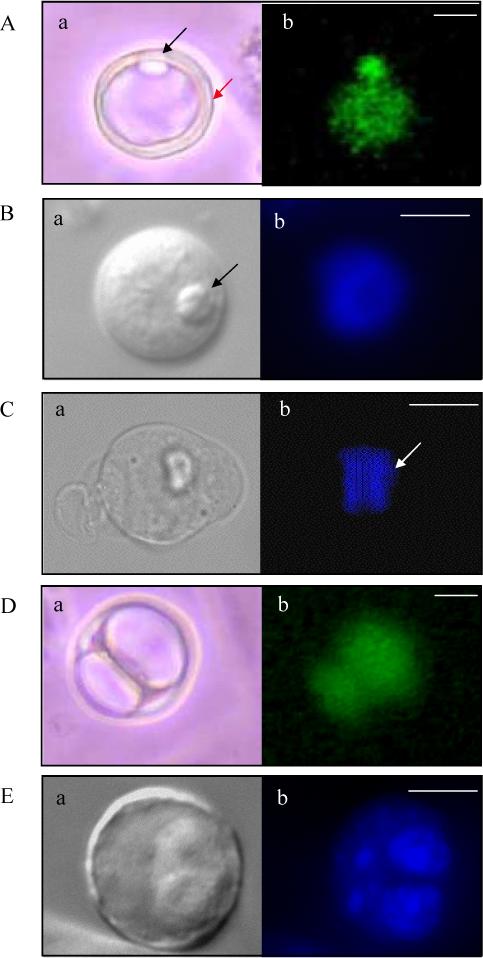
(A, B) eGFP-positive cells seen at d1 of in vitro differentiation of JM21 XX ES cells. Structures resembling polar bodies (black arrow) and a zona pellucida (red arrow) are seen. eGFP fluorescence represented in A, Hoechst DNA stain in B. (C) Hoechst stain of an oocyte-like cell at d1 of differentiation in metaphase stage of mitosis as determined by chromosomal alignment along a metaphase plate (white arrow). (D) eGFP positive zygote-like structure seen at d2 of in vitro differentiation and (E) Four-cell Hoechst stained cell seen at d2 of in vitro differentiation. (A-E, a): Phase images. Images in A and D = 20X magnification, Images in B, C and E = 40X magnification. Scale bars = 20μm (A, D), 25μm (B, C, E).