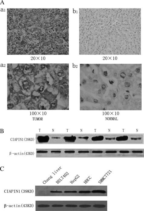
Fig. 1.
Immunohistochemical analysis of CIAPIN1 protein expression in non-tumor liver samples and HCC tissues (44 cases). (A) Representative photographs were taken at different magnifications (×200 versus ×1000 for inserts). (a1) HCC tissues ×200 magnification, (a2) HCC tissues ×1000 magnification; (b1) non-tumor liver samples ×200 magnification, (b2) non-tumor liver samples ×1000 magnification. (B) Western blot analysis of whole-cell protein extracts prepared from five paired non-tumor liver (N) and HCC (T) specimens. The level of CIAPIN1 protein expression was significantly higher in tumor tissue than in normal tissue. (C) CIAPIN1 protein expression is drastically increased in HCC cells compared with the normal human liver cells, Chang liver, determined by western blot analysis. β-actin expression levels were used as internal controls.

