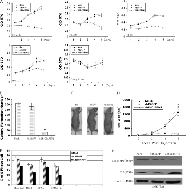
Fig. 3.
Adenovirus-delivered CIAPIN1 siRNA inhibits HCC cell growth, tumorigenesis and S-phase entry. (A) The growth of HCC cells and normal Chang liver infected with adenovirus was measured by 3-(4,5-dimethylthiazole-2-yl)-2,5-biphenyl tetrazolium bromide assay. Values were given at the indicated time points as the mean absorbance with a standard deviation of four wells. (B) Colony formation assay was performed by using SMMC-7721 cells infected with adenovirus for 96 h. The cells without any infection (mock) were used as control. The data were the averages from three independent triplicate experiments. *P < 0.05 relative to the mock group. (C) Ex vivo assay for the tumor suppression effect of AdSiCIAPIN1. SMMC-7721 cells were infected with adenoviruses or untreated cells (mock) in culture plates for 96 h. Then viable cells (3.0 × 10⁶) were injected subcutaneously into the dorsal flanks of five mice in each group. Photographs of a representative mouse in each group are shown. (D) Quantitation of tumor volume shown in (C). Tumor formation was scored weekly. Data were expressed as means ± standard deviation (n = 5). (E) AdSiCIAPIN1 decreased S-phase population in HCC cells after infection for 96 h. The S-phase population in the indicated cells was determined by flow cytometry and experiments were repeated three times. Error bars: standard deviation. (F) Cell cycle-related molecules were determined by western blot analysis. In the western blot analysis, depletion of CIAPIN1 expression could downregulate cyclinD1 but upregulate p27 expression in SMMC-7721-infected cells.

