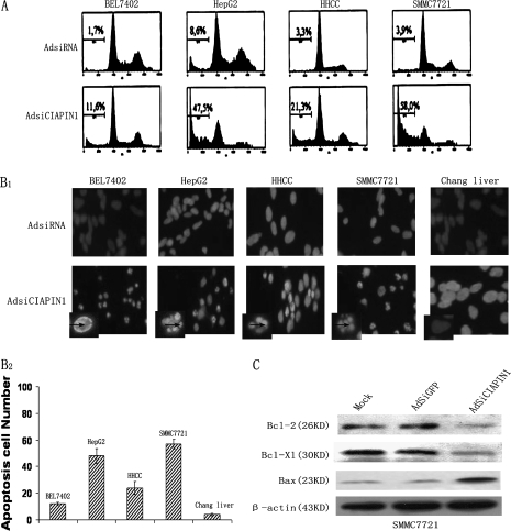
Fig. 4.
AdSiRNA-targeting CIAPIN1 induces cell apoptosis. (A) Downregulation of CIAPIN1 promoted apoptosis of the four indicated HCC cell lines. Ninety-six hours after the virus infection, the adherent cells were collected by trypsinization and determined by flow cytometry. Three individual experiments were performed, and the cell distribution in the cell cycle was determined by standard fluorescence-activated cell sorter analysis. The cell population in sub-G1 was shown. The x-axis and y-axis represent DNA content and the cell number, respectively. (B1) 4′,6-Diamidino-2-phenylindole staining showed the apoptotic cells in HCC cells infected with AdSiCIAPIN1 for 96 h, whereas this apoptosis cannot be seen in normal Chang liver being infected. The arrows indicate the blue color in apoptotic nuclei with condensed chromatin or nuclear membrane contiguousness morphology. (B2) Apoptosis cell numbers were counted in 100 cells under fluorescent microscopy, and results were representative of three independent experiments. (C) Western blot analysis of Bcl-2 family mediators of apoptosis showed that depletion of CIAPIN1 could downregulate Bcl-2 and Bcl-XL and upregulate Bax in AdSiCIAPIN1-transduced cells, suggesting that the apoptotic effect of CIAPIN1 could be partly mediated by the Bcl-2 family.

