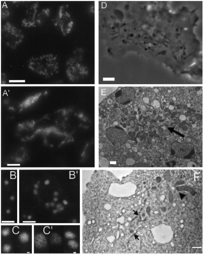
Figure 12.
Cells lacking a functional copy of dymA become multinucleated and display abnormal morphologies of nuclei and mitochondria. Panels A and A′ show cells stained with mAb 70-100-1 directed against a mitochondrial porin from D. discoideum. (A) Uniform, dispersed appearance of mitochondria in Ax2 cells. (A′) In dymA− cells dense clusters of mitochondria are frequently observed, and most mitochondria appear fused in a network of long threads. (B) Ax2 cells were stained with DAPI to visualize nuclei. (B′) DAPI staining of Dynamin A-depleted cells shows that these cells are multinucleated. Panels C and C′ show a gallery of DAPI-stained nuclei. In comparison to wild-type nuclei (C), dymA− nuclei (C′) display a lobed appearance and are more variable in size and shape. (D) The altered appearance of mitochondria in dymA− cells can also be observed in phase contrast images. (E) EM micrographs showing clustered mitochondria (arrow) in a dymA− cell. (F) EM micrographs showing a branched mitochondrial network consisting of thin filamentous structures (arrows) and large lobe-shaped structures (arrowhead) in dymA− cells. Bar, 10 μm in panels A, A′, B, and B′, and 1 μm in panels C, C′, D, E, and F.

