Figure 3.
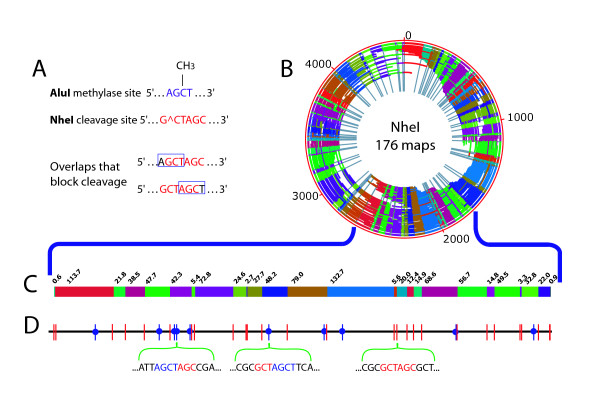
Profiling of E. coli AluI methylation sites by NheI restriction mapping. A; Overlaps between sites of NheI (red) restriction enzyme cleavage and AluI (blue) methylation blocking cleavage. B; NheI de novo optical map contig of AluI methylated E. coli, containing 176 maps. Outer red circle shows genome coordinates (kb) with internal arcs representing 176 maps; individual restriction fragments within each map are denoted by alternating colors; and grey radial lines demarcate restriction fragments within the contig map (next to the genome coordinates–red circle). The origin of the optical map does not coincide with the start of the published sequence, because the optical map was assembled de novo. C; An enlarged section (~960 kb) of the de novo NheI optical map contig. Colored blocks represent individual restriction fragments with their respective sizes (kb) marked above. D; Detailed comparison of the optical map shown in (C) against the corresponding in silico NheI map. NheI cleavage sites are shown as vertical bars; red bars show cleavage sites observed in the optical map. NheI sites overlapping with AluI methylation (absent in the optical map) are shown as blue vertical bars with a blue circle denoting blocking; below, the sequences around 3 NheI restriction sites are shown, two of which overlap AluI methylation sites.

