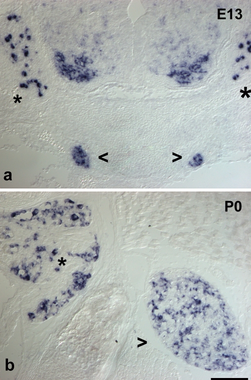
Fig. 2.
Expression of ret mRNA in sympathetic ganglia and DRG. In situ hybridization for ret mRNA on trunk cross sections from a 13-day-old mouse embryo (E13, a) and a newborn animal (P0, b). At E13, a population of large DRG (asterisks) neurons is positive, whereas many DRG cells are devoid of signal. Staining is found throughout the sympathetic ganglia (open arrowheads) albeit at various intensities. In newborn DRG, a small population of large neurons is strongly positive, whereas many small cells show weak signal. In sympathetic ganglia, a subset of cells is ret-positive at varying signal intensities. Bar 70 μm

