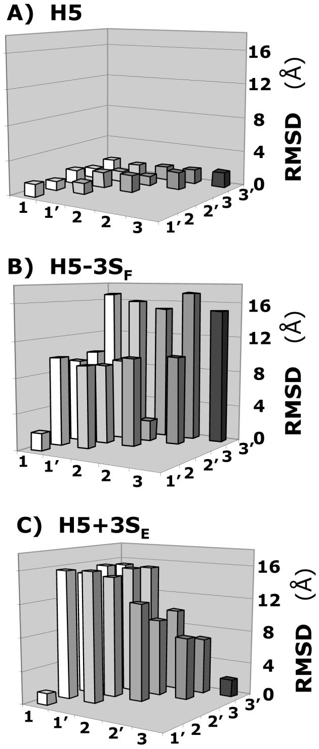
Figure 5. Specificity of GAG sequence for antithrombin.

Variation in the RMSD between six solutions (1, 1′, 2, 2′, 3, and 3′) obtained from three independent docking experiments of heparin pentasaccharide H5 variants, containing either one additional sulfate group at 3-position of residue E or lacking a sulfate at the 3-position of residue F, binding to antithrombin. Whereas for H5 (A) the RMSD was less than 2.0 Å between each solution, for H5-3SF (B) and H5+3SE (C), a majority of RMSD values were much greater (5-17 Å). Other pentasaccharide variants were also studied. See text for details.
