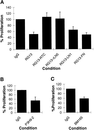
Figure 6.
(A) MCF-10A cells (2 × 10⁴ cells per well of a 24-well plate) were plated onto tissue culture plastic or surfaces coated with 50 μg/ml RTC, 2 μg/ml rtLN5, 25 μg/ml LN1, and 25 μg/ml FN for 48 hr. As indicated, MCF-10A cells were maintained in medium supplemented with either 50 μg/ml IgG control antibody or 50 μg/ml LN α3 subunit function-inhibiting antibody RG13. At 48 hr the cells were trypsinized and counted. % Proliferation indicates the increase in cell number as a percentage of that observed in the IgG-treated control cell population. The SD was determined from the data derived from three trials. At 48 hr the control cell population expanded from 2 × 10⁴ to 1 × 10⁵ cells (100%). (B) MCF-10A cells were maintained in medium supplemented with either 50 μg/ml IgG control antibody or 50 μg/ml LN5 function-inhibiting antibody P3H9-2. (C) MCF-10A cells were maintained in medium supplemented with either 50 μg/ml IgG control antibody or 50 μg/ml LN5 function-inhibiting antibody BM165. In B and C, the control cell populations expanded from 2 × 10⁴ to 1.1 × 10⁵ and from 2 × 10⁴ to 9.1 × 10⁴ after 48 hr (100%).

