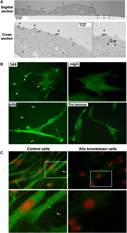
Figure 1.
Evidence that Alix is present in the extracellular compartment of WI38 cell cultures. (A) Monolayer cultures of WI38 cells were immunogold-labelled with 3A9 antibody, and electron micrographs of a sagittal section and a cross section of embedded samples are shown. Arrowheads indicate positive staining on the cell surface. Arrows indicate positive staining on the substratum. (B) Monolayer cultures of WI38 cells fixed with the EM fixative were immunostained under identical conditions with 3A9 antibody, mouse IgG (mIgG), rabbit anti-Alix immune serum (pAb) or pre-immune serum (pre-immune). Arrows indicate positive staining on the substratum and at the cell periphery. (C) Monolayer cultures of control and Alix-knockdown WI38 cells fixed with methanol were immunostained with 3A9 antibody (green) and counterstained with PI (red). Arrows indicate positive staining on the substratum.

