Figure 3.
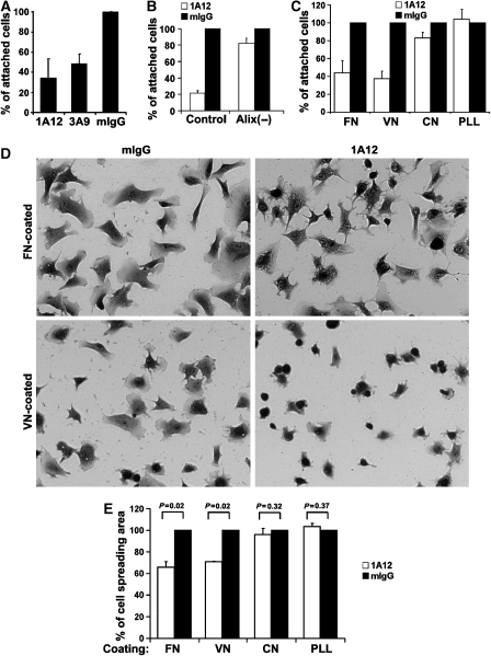
Anti-Alix antibodies inhibit integrin-mediated cell adhesions. (A) WI38 cells were seeded in the presence of each of the indicated antibodies, and relative cell attachments were determined at 1 h after cell seeding. Results were normalized against the value from mouse IgG (mIgG)-treated cells, and presented results are averages from three independent experiments. Error bars indicate standard errors of mean (s.e.m.). (B) Control and Alix-knockdown WI38 cells were seeded in the presence of 1A12 antibody or mIgG, and relative cell attachments were determined at 1 h after cell seeding. Presented results are averages from three independent experiments, and the error bars indicate standard errors of mean (s.e.m.). (C) WI38 cells were seeded onto the substratum that was pre-coated with FN, vitronectin (VN), collagen (CN) or poly-L-lysine (PLL) in the presence of 1A12 antibody or mIgG, and relative cell attachments on each of the coated proteins were determined at 1 h as described for (A). Results are from a representative experiment out of three, and error bars indicate standard deviations. (D, E) WI38 cells were seeded as described for (C), and cells were stained with crystal violet at 2 h after cell seeding and photographed (D). The relative spreading area per cell was determined by analysis of digitized images with Metamorph software, and the average was calculated and normalized against the value of mIgG-treated cells (E). The P-values were determined using Student's t-tests.

