Figure 3.
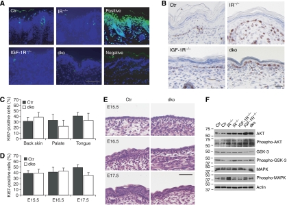
Apoptosis and proliferation in the epidermis in the absence of IR/IGF-1R. (A) TUNEL staining (green) on sections isolated from back skin of newborn mice. Nuclei were counterstained using DAPI (blue). Scale bar is 100 μm. (B) Ki67 staining on sections of back skin isolated from newborn mice. Scale bar is 100 μm. (C) Quantification of Ki67 staining in the basal cells of IFE of control and dko mice in back skin, palate or tongue epithelium. N=5 and P>0.05 for each genotype. (D) Quantification of Ki67 staining of the epidermis in embryos. N=4 mice/group, P>0.05 for E15.5 and 16.5, P<0.01 for E17.5 (E) H&E staining on paraffin sections from E15.5, E16.5 and E17.5 embryos. Scale bar is 50 μm. (F) Western blot analysis for the indicated proteins on epidermal lysates of newborn mice.

