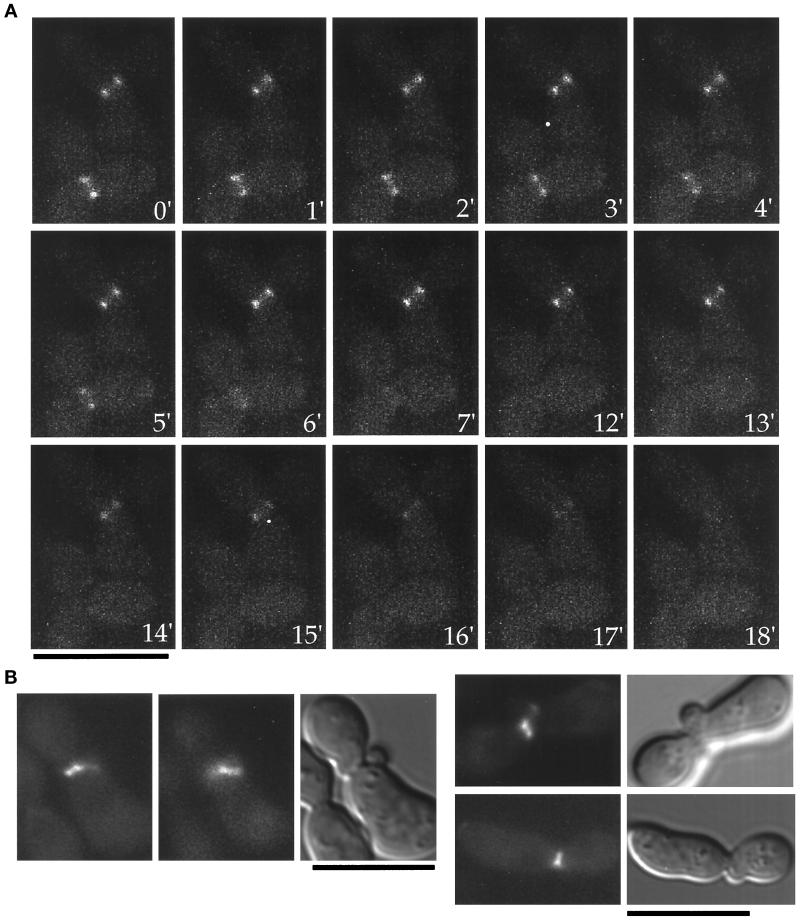
Figure 2.
Myo1-GFP in Cyk1− cells. (A) A representative time lapse sequence of the Myo1-GFP ring after GAL1 promoter shut off of Cyk1p expression. (B) An example of a Cyk1p− cell with Myo1-GFP located at both old and new bud necks in a cell. Two focal planes of Myo1-GFP are shown, showing old (left) and new (right) rings. (C) Examples of Cyk1p− cells with Myo1-GFP remaining at old bud neck and failing to form a ring at the new neck. Bars, 10 μm.

