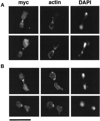
Figure 4.
Effects of ΔCHD (A) and ΔGRD (B) on the formation of the Cyk1p (myc) and actin rings. Cells were fixed and stained with anti-myc primary, FITC-conjugated anti-mouse secondary antibody (left), rhodamine phallodin (middle), and DAPI (right). Representative cells that have a myc ring are shown. Bar, 10 μm.

