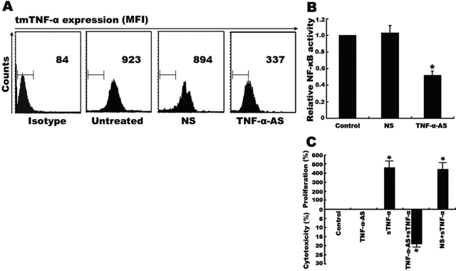
Fig. 4.
Constitutive NF-κB activation and resistance to sTNF-α cytotoxicity mediated by overexpression of endogenous TNF-α. Raji cells (2×10⁶) were transfected with 10 μM TNF-α antisense or with 10 μM mismatched NS for 24 h. (A) tmTNF-α expression on the surface of Raji cells was detected by flow cytometry using a polyclonal antibody specific to TNF-α. The results as shown by mean fluorescence intensity (MFI) are representative of at least three independent experiments. (B) The transfectants and the nontransfected cells were lysed, and the NF-κB activity was measured by ELISA. (C) Cells (5×10⁴) with transfection of TNF-AS or NS or with nontransfection were incubated with 100 U/ml sTNF-α for 24 h. The viability of cells was determined by MTT. Data represent the mean of three independent experiments; *, P < 0.01.

