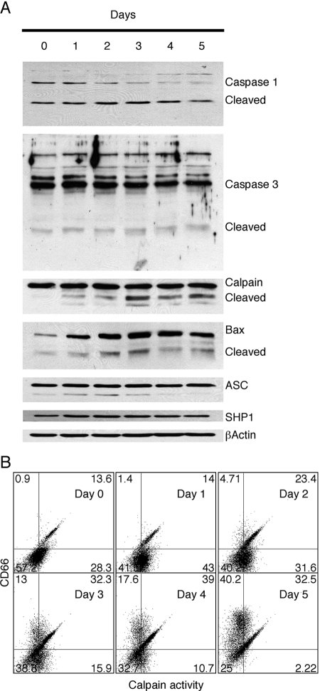
Fig. 5.
Analysis of ATRA-treated HL-60 cell for markers of apoptosis. (A) HL-60 cells were treated with 2 μM ATRA for 0–5 days and analyzed by Western blot analysis for expression of caspase-1, caspase-3, calpain, Bax, ASC, and SHP1. In each case, note the presence or absence of the cleaved, activated form of the proapoptotic protein. (B) FACS analysis of ATRA-treated cells was performed by double-staining with the calpain substrate rhodamine 110, bis-(t-BOC-L-leucyl-L-methionine amide), and CD66 antibody T84.1 All experiments were repeated at least twice, and similar results were obtained.

