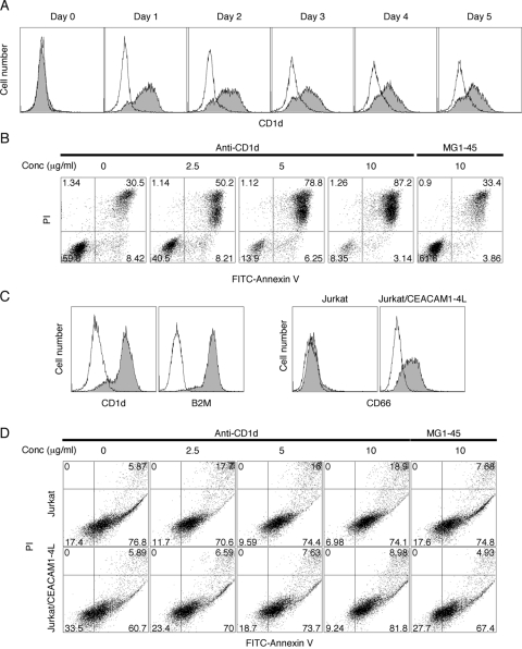
Fig. 6.
CD1d induction and apoptosis with its stimulation on leukemic cell lines. HL-60 cells were treated with 2 μM ATRA for 0–5 days, continuously or transiently. (A) CD1d expression (shaded) was determined by FACS. The isotype control is shown (open). (B) ATRA-treated HL-60 cells were stimulated with anti-CD1d antibody at Day 1 for an additional 2 days. Apoptosis was analyzed by FITC-annexin V/PI double-staining. MG1-45 isotype antibody was used as negative control. (C) CD1d, B2M, and CD66 expressions in Jurkat cells were measured by FACS. (D) Jurkat cells transfected with or without CEACAM1-4L were stimulated with anti-CD1d antibody for 2 days, and apoptosis was analyzed with FITC-annexin V/PI double-staining. All experiments were repeated at least twice with consistent results.

