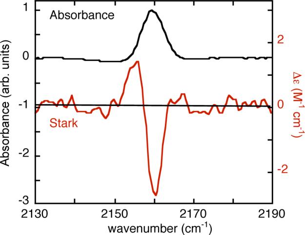
FIGURE 1.

IR absorption (top, left axis) and VSE (red, bottom, right axis) spectra for 11 mM RNase S with homocysteine introduced at position 13 and labeled with CN. The VSE spectrum is the field-on minus field-off difference spectrum obtained at 77K in a 50/50 (v/v) glycerol/water glass. The spectrum is scaled to a pathlength of 1 cm and a field of 1 MV/cm (actual field was 0.67 MV/cm).
