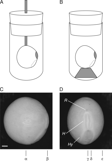
Fig. 1.
Soybean set in a sample tube for the acquisition of MR images. Two methods were employed: (A) a soybean was fixed by a plastic binder to a wooden stick attached to the lid of a 15-mm test tube, and (B) a soybean was fixed by a plastic binder to a small bed placed at the bottom of the test tube. The positions of sliced sections of images are indicated in (C) and (D): sections are at α in Figs 2, 4 and 9, and at γ in Figs 3, 6A–F and 7A–G; the sections used for MIP images are from δ to ε in Figs 6 G–L and 7H–N, and from α to β in Fig. 8. Abbreviations: R, raphe; H, hilum; Hy, hypocotyle–radicle axis. Scale bar in (C) = 1 mm.

