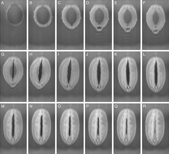
Fig. 2.
Changes in a soybean during imbibition at a median longitudinal section (Fig. 1C, α) normal to the raphe–antiraphe of the longer axis. A soybean (‘Mikawashima’) was fixed by the method indicated in Fig. 1A. Images were acquired continuously for 20 h at 15-min intervals after 5 min of imbibition, and those presented here are at 60-min intervals from 5 min of imbibition for a period of 17 h, as follows: (A) 5 min, (B) 1 h 5 min, (C) 2 h 5 min, (D) 3 h 5 min, (E) 4 h 5 min, (F) 5 h 5 min, (G) 6 h 5 min, (H) 7 h 5 min, (I) 8 h 5 min, (J) 9 h 5 min, (K) 10 h 5 min, (L) 11 h 5 min, (M) 12 h 5 min, (N) 13 h 5 min, (O) 14 h 5 min, (P) 15 h 5 min, (Q) 16 h 5 min, and (R) 17 h 5 min. Highlighted signals represent water taken up.

