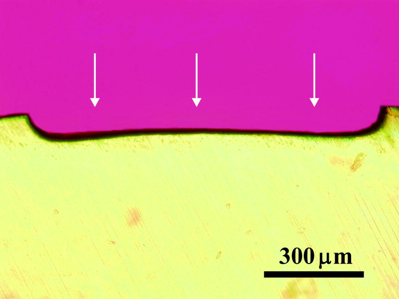
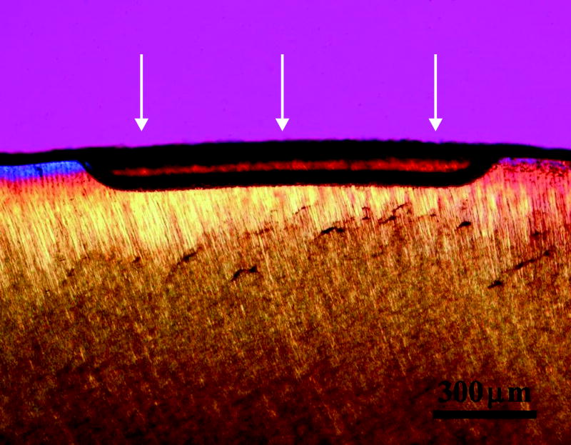
Figure 1.
Enamel or root surfaces were painted with acrylic nail polish to expose windows. The teeth were suspended for 25 hours in Coke®, which was replaced every 5 hours. The teeth were sectioned perpendicular to windows. Sections were viewed using a polarized light microscope and photographed. Representative lesions (arrows) produced in enamel (a) and root (b) surfaces are shown.


