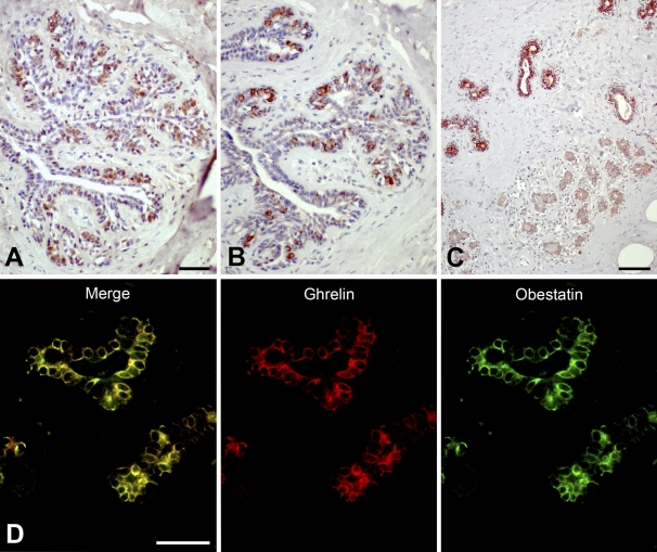
Figure 4.
Immunostaining for obestatin and ghrelin peptides in human mammary glands. Ghrelin (A) and obestatin (B) immunoreactivity is detected in inner cells of the ducts. Occasional weaker staining is found in the lobuli. (C) Section taken from inner part of the glands showing obestatin immunoreactivity in the ductal epithelium, whereas occasional immunoreactive cells are found in the lobuli. (D) Immunofluorescence staining of ducts in mammary tissue. Ghrelin [tetramethyl rhodamine isothiocyanate (TRITC)] is visualized as red and obestatin (FITC) as green. Yellow color in the merged image indicates co-localization of obestatin and ghrelin. Bars: A,B = 50 μm; C = 100 μm; D = 20 μm.

