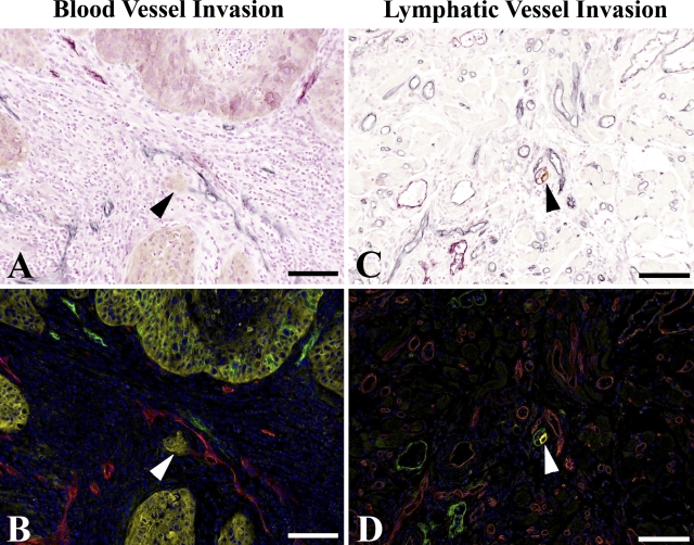
Figure 2.
Triple-stain immunohistochemistry and multispectral imaging identify occurrences of vessel invasion. Intratumoral invasion of tumor cells into blood vessel (A,B), arrowheads. Peritumoral invasion of tumor cell into lymphatic vessel (C,D), arrowheads. Triple-stain method under light microscope shows blood vessels as blue and lymphatic vessels as purple (A,C); unmixing by multispectral analysis shows blood vessels as red and lymphatic vessels as green (B,D). Blood and lymphatic vessels are clearly distinguished. Bar = 0.1 mm.

