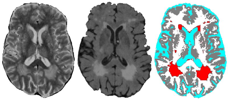
Figure 1. Brain lesions seen on magnetic resonance imaging (MRI)1.

1Lesions, believed to be of ischemic origin, occur with aging but particularly with late-life depression. These lesions may damage mood regulation pathways (leading to depression) or cause cognitive impairment. Lesions are shown here on a proton density image (left), fluid-attenuated inversion recovery (FLAIR) image (center), and tissue classification image (right). Lesions are red on tissue classification image. Images are courtesy of the Neuropsychiatric Imaging Research Laboratory at Duke University.
