
Fig. 2.
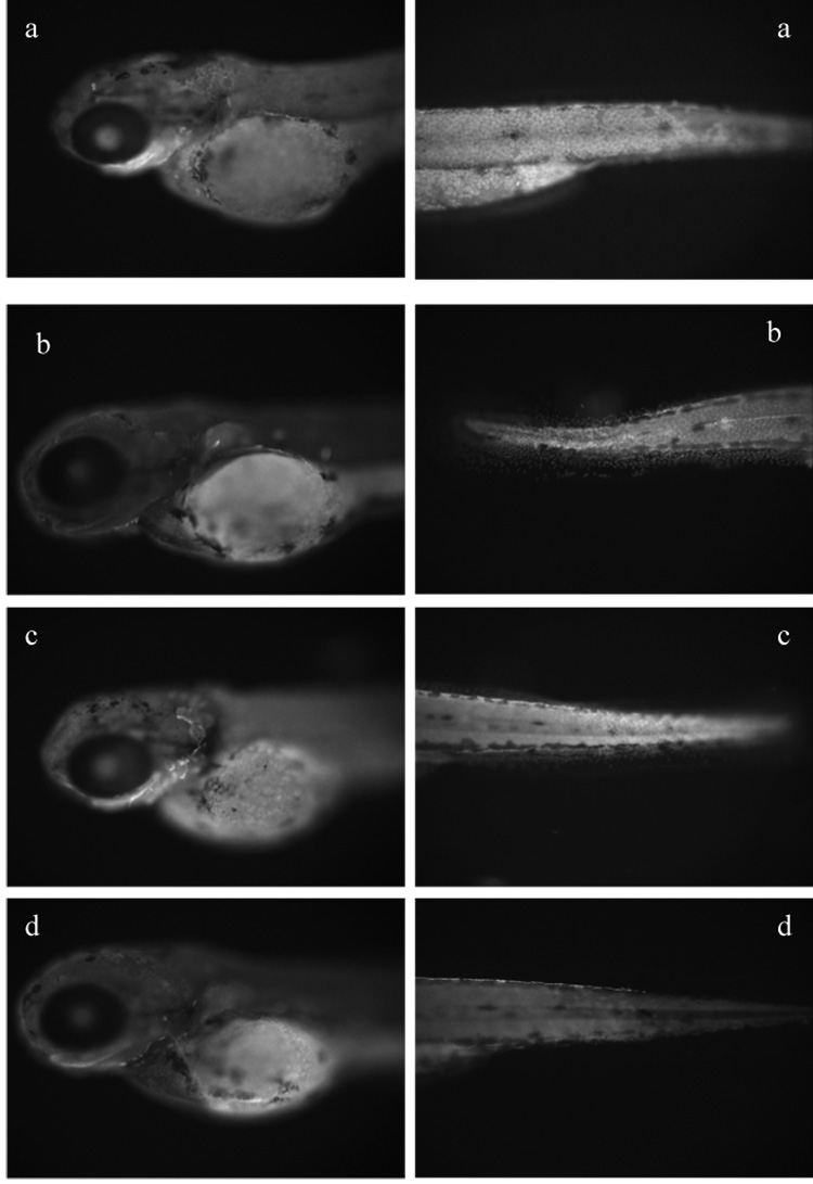
Immunolocalization of CYP1B1 (530 nm excitation and 586 nm emission filter; 100X, f500 microscopy). (A) Control-morpholino embryo exposed to PCB 126 showing induction of CYP1B1 indicated by strong fluorescence in epithelial cells. (B) Control-morpholino embryo exposed to DMSO showing less CYP1B1 induction, as indicated by limited fluorescence compared to (A). (C) CYP1B1 morpholino embryo exposed to DMSO and (D) CYP1B1 morpholino embryo exposed to PCB 126 showing less CYP1B1 induction as indicated by limited fluorescence compared to (A).